


With the osteochondrosis of the spine, many are not familiar with the popular television screen, but of their own sad experience.The statistics are hard: up to 80% of the population suffers from this ailment, which is also significantly younger.If the previous complaints about problems in the spine were mainly among the previous generation, now the osteochondrosis of children no longer surprises anyone.And the fault of a sedentary lifestyle and the "benefits of civilization."

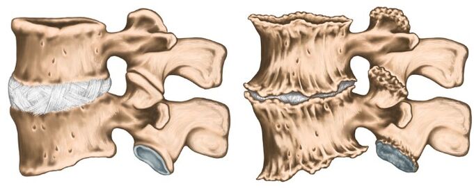
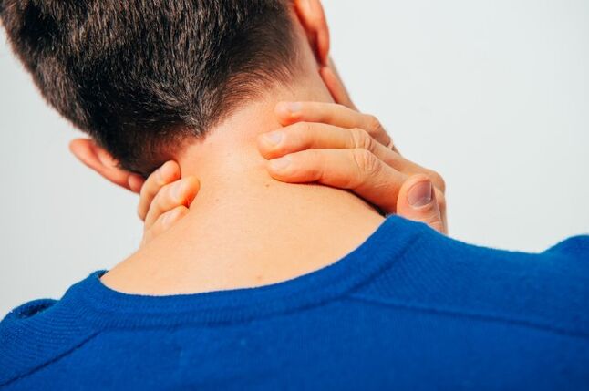
The osteochondrosis of the cervical column is polyietiological a progressive disease that is manifested by the degeneration of the intervertebral discs and the dystrophy of the spine ligament apparatus.Everyone knows first -hand symptoms, but this knowledge is fragmentary;We will try to structure them, as well as talk about the principles of diagnosis and the treatment of the osteochondrosis of the cervical column.
The causes of osteochondrosis
Medical science cannot respond unequivocally, so osteochondrosis occurs.It is known reliably that the sedentary lifestyle is that a modern person is prone to negatively affect the progression of this disease.It is interesting that both hypodinamia and colossal athlete loads lead to the proxy of the discs.A hereditary factor plays a main role.The following reasons are distinguished:
- loaded hereditary history;
- obesity;
- hypodinamia;
- metabolic disorders in the body;
- traumatic damage to the spinal column;
- Long static overloads and work associated with weightlifting (computer work, weightlifting, miners, engines, etc.);
- scoliosis;
- dysfunctional environmental situation;
- flat feet and pregnancy;
- hypothermia and stress, which often cause exacerbations of the disease.
There are several neurological syndromes:
- shoulder periartritis;
- root;
- cardiac;
- Vail artery syndrome.
Shoule -Houlder Periartritis.It is characterized by neck pain, shoulder, shoulder joint.The leading neurogenic contracture of the shoulder joint is formed, which is of a protective nature, since it protects the axillary nerve from the stretching (it is antalgic).With this position, the muscles surrounding the joint are in tension.The severity of pain syndrome depends on the exacerbation of osteochondrosis: in a slight limitation of the amplitude of the articulation movements to the "frozen shoulder" called SO, when any movement causes severe pain.The pain intensifies when the shoulder deviates and pronounces, since it is these movements that improve the voltage of the axillary nerve.
Royshift Syndrome (Cervical Radiculita).Most of the time it happens with cervical osteochondrosis.At the same time, the spinal spinal spinal is squeezed due to the "sinking" of the intervertebral discs, as well as due to the growth of osteofites or the protuberance of the discs in the lateral direction.The pain syndrome is specific: intense burning, tear, pressing pain, which also intensifies when the patient moves the head.The antalgic pose is also observed in the neck muscles, they are very tense and painful, the volume of movements is limited.There is pain on the back of the head, neck, front chest, shoulder, among the shoulder blades.The interruption of sensitivity by the type of "jacket with short sleeves" is characteristic.
Cardial syndrome.The name of the syndrome is responsible for itself: the clinical image is very similar to the angina pectoris.In this case, there is no organic damage in the heart, at the height of the pain syndrome, violations of coronary blood flow by ECG are not detected, and such patients are well tolerated.A typical characteristic with angina pectoris: pain takes place after taking nitrates, and in the case of osteochondrosis does not change and bothers for a long time.Unlike angina pectoralis, pain location is mainly in the heart of the left.With irritation of the roots of segments C8 - T1, tachycardia and extrastole rhythm disturbances are possible.This is not due to the damage to the heart driving system, but to a violation of the sympathetic innervation of the heart muscle (extracardiac damage).In the differential diagnosis of angina pectoris and cardiac syndrome, leadership is the fact that, in addition to cardial complaints, the patient indicates the increase in pain in the shoulder and neck joint associated with lifting or hard movements.
Vail artery syndrome.The vertebral artery takes place in a channel formed by holes in the transverse processes of the vertebrae.This artery is matched, is responsible for the supply of blood to the brain.Consequently, any narrowing of this channel affects the nutrition of brain tissue very negatively.The vertebral artery syndrome develops directly both with the compression of the artery itself as with the irritation of the sympathetic nerve plexus, which is around it.The pain in this pathology is burning or pressing in the occipital region with whiskey propagation, tutorial arches, crows.It arises both on one and both sides.Patients generally associate aggravation with the condition after sleep in a non -physiological pose, travel travel, walk.With pronounced symptoms, hearing loss, dizziness, noise in the ears, nausea, vomiting, loss of consciousness and increased blood pressure.Such symptoms are not specific and are very similar to complaints in brain stroke.This pathology is characterized by the syndrome of the Sistine Chapel: a fainting that occurs when you revoke your head (severe cerebral ischemia).The visitors of the Sistine Chapel described it in the Vatican when they examined the frescoes in their arches.It is also possible to fall without loss of consciousness with acute head turns.
Like any diagnosis in medicine, the diagnosis of osteochondrosis is established on the basis of patient complaints, disease anamnesis, clinical examination and auxiliary research methods.X -rays of the cervical column are performed in direct and lateral projections, if necessary in special positions (with an open mouth).At the same time, experts are interested in the height of the intervertebral discs, the presence of osteofites.From modern research methods, IAMR and CT research is used, which allows to verify the diagnosis with greater precision.In addition to additional listing methods, consultations of related specialists (cardiologist, ophthalmologist, neurosurgeon) may be necessary, and neurologist's exam is simply vital.The neurologist is dedicated to the treatment of osteochondrosis, so after examining the patient, he will prescribe the minimum necessary examination to his discretion.
Osteochondrosis treatment
Osteochondrosis is a poleetiological disease, since a therapy course is not cured.You cannot drink a "magic pill" and everything will happen, it is necessary to fundamentally change your lifestyle, since the trigger is hypodinamia.The most tangible results are easier to achieve in the initial stage of the disease, when complaints are minimal and there are no compression syndromes and spinal artery.In the acute stage of the disease, when the following groups of medicines are prescribed that pronounced pain: pain syndrome is pronounced:
- Therapeutic therapy block (to relieve pain and elimination of muscle spasm);
- NSAID;
- Ointments containing NSAIDs and action reflects;
- muscle relaxants;
- B Vitamins V.
As the inflammatory process decreases and the relief of pain syndrome, they go to physiotherapy treatment.Very often, the following techniques are used:
- laser therapy;
- electrophoresis;
- acupuncture;
- Exercise therapy;
- therapeutic massage;
- Manual therapy
It is important to understand that osteochondrosis continues with periods of exacerbation and remission, therefore, it is very important to affect the cause and not treat research.